
File:Blood cell crossing vascular sinus wall - TEM.jpg
Size of this preview: 597 × 480 pixels.
| |
This is a file from the Wikimedia Commons. Information from its description page there is shown below.
Commons is a freely licensed media file repository. You can help. |
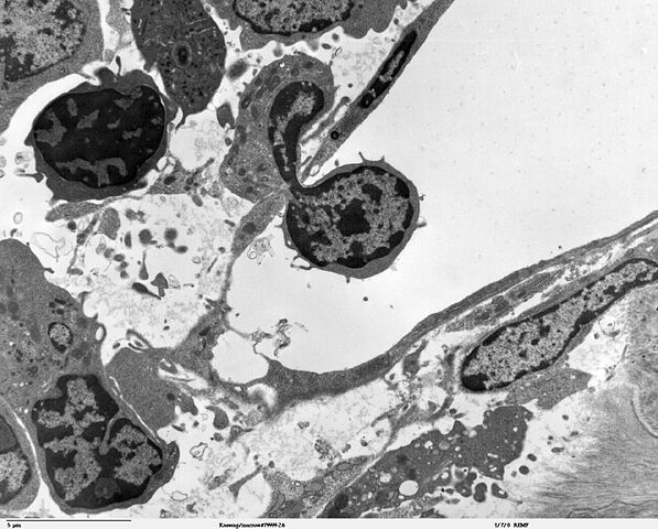
| Description | Transmission electron micrscope image of a thin section cut through an area of bone marrow area near the cartilage/bone interface in a mouse kneecap. Image shows small opening in the thin endotheliun of the vascular sinus wall, where a blood cell is crossing the thin vascular sinus wall and into the sinus lumen. JEOL 100CX TEM |
| Date | |
| Source |
|
| Author | Louisa Howard, Roy Fava |
| Permission ( Reusing this file) |
PD |
Licensing
 |
This work has been released into the public domain by its author, Louisa Howard and Roy Fava. This applies worldwide. In some countries this may not be legally possible; if so: Louisa Howard and Roy Fava grants anyone the right to use this work for any purpose, without any conditions, unless such conditions are required by law.
|
File usage
The following pages on Schools Wikipedia link to this image (list may be incomplete):
About Schools Wikipedia
SOS Childrens Villages has brought Wikipedia to the classroom. Thanks to SOS Children's Villages, 62,000 children are enjoying a happy childhood, with a healthy, prosperous future ahead of them. Sponsoring a child is the coolest way to help.