Optical microscope
Background to the schools Wikipedia
SOS Children, an education charity, organised this selection. With SOS Children you can choose to sponsor children in over a hundred countries

The optical microscope, often referred to as the "light microscope", is a type of microscope which uses visible light and a system of lenses to magnify images of small samples. Optical microscopes are the oldest and simplest of the microscopes.
There are non-optical microscopes, which require chemical or ion staining of non-living samples, and can magnify exponentially greater than the optical microscope. See: scanning electron microscope, transmission electron microscope.
Optical configurations
There are two basic configurations of optical microscope in use, the simple (one lens) and compound (many lenses).
Simple optical microscope
A simple microscope is a microscope that uses only one lens for magnification, and is the original light microscope. Van Leeuwenhoek's microscopes consisted of a small, single convex lens mounted on a brass plate, with a screw mechanism to hold the sample or specimen to be examined. Demonstrations by British microscopist have produced surprisingly detailed images from such basic instruments. Though now considered primitive, the use of a single, convex lens for viewing is still found in simple magnification devices, such as the magnifying glass, and the loupe. Light microscope are able to view specimens in colour, an important advantage when compared with electron microscopes, especially for forensic analysis.
History of the microscope
It is difficult to say who invented the compound microscope. Dutch spectacle-makers Hans Janssen and his son Zacharias Janssen are often said to have invented the first compound microscope in 1590, but this was a declaration made by Zacharias Janssen himself during the mid 1600s. The date is unlikely, as it has been shown that Zacharias Janssen actually was born around 1590. Another favorite for the title of 'inventor of the microscope' was Galileo Galilei. He developed an occhiolino or compound microscope with a convex and a concave lens in 1609. Galileo's microscope was celebrated in the Accademia dei Lincei in 1624 and was the first such device to be given the name "microscope" a year latter by fellow Lincean Giovanni Faber. Faber coined the name from the Greek words μικρόν (micron) meaning "small", and σκοπεῖν (skopein) "meaning to look at", a name meant to be analogus with "telescope", another word coined by the Linceans.
Christiaan Huygens, another Dutchman, developed a simple 2-lens ocular system in the late 1600s that was achromatically corrected, and therefore a huge step forward in microscope development. The Huygens ocular is still being produced to this day, but suffers from a small field size, and other minor problems.
Anton van Leeuwenhoek (1632-1723) is generally credited with bringing the microscope to the attention of biologists, even though simple magnifying lenses were already being produced in the 1500s. Van Leeuwenhoek's home-made microscopes were very small simple instruments, with a single, yet strong lens. They were awkward in use, but enabled van Leeuwenhoek to see detailed images. It took about 150 years of optical development before the compound microscope was able to provide the same quality image as van Leeuwenhoek's simple microscopes, due to timely difficulties of configuring multiple lenses. Still, despite widespread claims, van Leeuwenhoek is not the inventor of the microscope.
The components of the microscope
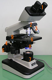
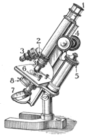
All optical microscopes share the same basic components:
- The eyepiece - a cylinder containing two or more lenses to bring the image to focus for the eye. The eyepiece is inserted into the top end of the body tube. Eyepieces are interchangeable and many different eyepieces can be inserted with different degrees of magnification. Typical magnification values for eyepieces include 5x, 10x and 2x. In some high performance microscopes, the optical configuration of the objective lens and eyepiece are matched to give the best possible optical performance. This occurs most commonly with apochromatic objectives.
- The objective lens - a cylinder containing one or more lenses to collect light from the sample. At the lower end of the microscope tube one or more objective lenses are screwed into a circular nose piece which may be rotated to select the required objective lens. Typical magnification values of objective lenses are 4x, 5x, 10x, 20x, 40x, 80x and 100x. Some high performance objective lenses may require matched eyepieces to deliver the best optical performance.
- The stage - a platform below the objective which supports the specimen being viewed. In the centre of the stage is a circular hole through which light passes to illuminate the specimen. The stage usually has arms to hold slides (rectangular glass plates with typical dimensions of 25 mm by 75 mm, on which the specimen is mounted).
- The illumination source - below the stage, light is provided and controlled in a variety of ways. At its simplest, daylight is directed via a mirror. Most microscopes, however, have their own controllable light source that is focused through an optical device called a condenser, with diaphragms and filters available to manage the quality and intensity of the light.
The whole of the optical assembly is attached to a rigid arm which in turn is attached to a robust U shaped foot to provide the necessary rigidity. The arm is usually able to pivot on its joint with the foot to allow the viewing angle to be adjusted. Mounted on the arm are controls for focusing, typically a large knurled wheel to adjust coarse focus, together with a smaller knurled wheel to control fine focus.
Updated microscopes may have many more features, including transmission illumination, phase contrast microscopy and differential interference contrast microscopy, and digital cameras.
On a standard compound optical microscope, there are three objective lenses: a scanning lens (4×), low power lens (10×)and high power lens (40×). Advanced microscopes often have a fourth objective lens, called an oil immersion lens. To use this lens, a drop of immersion oil is placed on top of the cover slip, and the lens is very carefully lowered until the front objective element is immersed in the oil film. Such immersion lenses are designed so that the refractive index of the oil and of the cover slip are closely matched so that the light is transmitted from the specimen to the outer face of the objective lens with minimal refraction. An oil immersion lens usually has a power of 100×.
The actual power or magnification of an optical microscope is the product of the powers of the ocular ( eyepiece), usually about 10×, and the objective lens being used.
Compound optical microscopes can produce a magnified image of a specimen up to 1000× and, at high magnifications, are used to study thin specimens as they have a very limited depth of field.
How a microscope works
The optical components of a modern microscope are very complex and for a microscope to work well, the whole optical path has to be very accurately set up and controlled. Despite this, the basic optical principles of a microscope are quite simple.
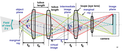
The objective lens is, at its simplest, a very high powered magnifying glass i.e. a lens with a very short focal length. This is brought very close to the specimen being examined so that the light from the specimen comes to a focus about 160 mm inside the microscope tube. This creates an enlarged image of the subject. This image is inverted and can be seen by removing the eyepiece and placing a piece of tracing paper over the end of the tube. By carefully focusing a rather dim specimen, a highly enlarged image can be seen. It is this real image that is viewed by the eyepiece lens that provides further enlargement.
In most microscopes, the eyepiece is a compound lens, with one component lens near the front and one near the back of the eyepiece tube. This forms an air-separated couplet. In many designs, the virtual image comes to a focus between the two lenses of the eyepiece, the first lens bringing the real image to a focus and the second lens enabling the eye to focus on the virtual image.
In all microscopes the image is viewed with the eyes focused at infinity (mind that the position of the eye in the above figure is determined by the eye's focus). Headaches and tired eyes after using a microscope are usually signs that the eye is being forced to focus at a close distance rather than at infinity.
Stereo microscope
The stereo or dissecting microscope is designed differently from the diagrams above, and serves a different purpose. It uses two separate optical paths with two objectives and two eyepieces to provide slightly different viewing angles to the left and right eyes. In this way it produces a three-dimensional visualization of the sample being examined.
The stereo microscope is often used to study the surfaces of solid specimens or to carry out close work such as sorting, dissection, microsurgery, watch-making, small circuit board manufacture or inspection, and the like.
Unlike compound microscopes, illumination in a stereo microscope most often uses reflected (episcopic) illumination rather than transmitted (diascopic) illumination, that is, light reflected from the surface of an object rather than light transmitted through an object. Use of reflected light from the object allows examination of specimens that would be too thick or otherwise opaque for compound microscopy. However, stereo microscopes are also capable of transmitted light illumination as well, typically by having a bulb or mirror beneath a transparent stage underneath the object, though unlike a compound microscope, transmitted illumination is not focused through a condenser in most systems. Stereoscopes with specially-equipped illuminators can be used for dark field microscopy, using either reflected or transmitted light.
Great working distance and depth of field here are important qualities for this type of microscope. Both qualities are inversely correlated with resolution: the higher the resolution (i.e. the shorter the distance at which two adjacent points can be distinguished as separate), the smaller the depth of field and working distance. A stereo microscope has a useful magnification up to 100×. The resolution is maximally in the order of an average 10× objective in a compound microscope, and often much lower.
There are two types of magnification systems in stereo microscopes. One is fixed magnification in which primary magnification is achieved by a paired set of objective lenses with a set degree of magnification. The other is zoom or pancratic magnification, which are capable of a continuously variable degree of magnification across a set range. Zoom systems can achieve further magnification through the use of auxiliary objectives that increase total magnification by a set factor. Also, total magnification in both fixed and zoom systems can be varied by changing eyepieces.
The stereo microscope should not be confused with a compound microscope equipped with binocular eyepieces. In such a microscope both eyes see the same image, but the binocular eyepieces provide greater viewing comfort. However, the image in such a microscope is no different from that obtained with a single monocular eyepiece.
Digital display with stereo microscopes
Recently various video dual CCD camera pickups have been fitted to stereo microscopes, allowing the images to be displayed on a high resolution LCD monitor. Software converts the two images to an integrated Anachrome 3D image, for viewing with plastic red/cyan glasses, or to the cross converged process for clear glasses and somewhat better colour accuracy. The results are viewable by a group wearing the glasses. These files may recorded as well.
Special designs
Other types of optical microscope include:
- the inverted microscope for studying samples from below; useful for cell cultures in liquid;
- the student microscope designed for low cost, durability, and ease of use;
- the research microscope which is an expensive tool with many enhancements;
- the petrographic microscope whose design usually includes a polarizing filter, rotating stage and gypsum plate to facilitate the study of minerals or other crystalline materials whose optical properties can vary with orientation.
- the polarizing microscope
- the fluorescence microscope
- the phase contrast microscope
Limitations of light microscopes
At very high magnifications with transmitted light, point objects are seen as fuzzy discs surrounded by diffraction rings. These are called Airy disks. The limit of resolution is therefore taken as the ability to distinguish between two closely spaced Airy disks. It is these impacts of diffraction that limit the ability to resolve fine details. The extent of and magnitude of the diffraction patterns are affected by both by the wavelength of light ( ), the refractive materials used to manufacture the objective lens and the numerical aperture (NA or
  ) of the objective lens. There is therefore a finite limit beyond which it is impossible to resolve separate points in the objective field. Assuming that optical aberrations in the whole optical set-up are negligible, the resolution d, is given by:
Usually, a  of 550 nm is assumed, corresponding to green light. With air as medium, the highest practical
  is 0.95, and with oil, up to 1.5. In practice the lowest value of d obtainable is around 0.2 micrometres or 200 nanometers.

Other optical microscope designs (e.g. Stimulated Emission Depletion Microscopy) can offer an improved resolution when observing self-luminous particles, which is not covered by Abbe's diffraction limit for the compound microscope. Abbe's theory (by Ernst Karl Abbe) is based on the fact that a non-self-luminous particle is illuminated by an extraneous source. For Ernst Abbe's work in light microscopy, see the Molecular Expressions web site at http://micro.magnet.fsu.edu/optics/timeline/people/abbe.html.
Alternatives to optical microscopy
In order to overcome the limitations set by the diffraction limit of visible light other microscopes have been designed which use other waves.
- Transmission electron microscope
- Scanning electron microscope
- X-ray microscope
The use of electrons and x-rays in place of light allows much higher resolution - the wavelength of the radiation is shorter so the diffraction limit is lower. To make the short-wavelength probe non-destructive, the atomic beam imaging system ( atomic nanoscope) is proposed and widely discussed in the literature, but it is not yet competitive with conventional imaging systems.


